02 / EVIDENCE
The thymosin alpha-1 clinical trial record, read closely.
From the 1977 first isolation through the 2025 TESTS phase 3 null result — every major study, described precisely.
The short version
This page walks what the clinical trials actually found, from the 1977 first isolation through the 2025 phase-3 sepsis null result. The hepatitis B and C data is the most consistent — one key trial found a 48.3% complete response rate versus 27.3% for interferon-alpha [3]. The sepsis data is contested: earlier trials showed a roughly nine-point mortality reduction; the largest phase-3 trial (1,106 patients, double-blind) found no effect [16]. The cancer data is intriguing but entirely retrospective. The mechanism behind all of it — binding receptors TLR2 and TLR9 on dendritic cells, which then mature T cells — is well characterized. What the evidence base does not support: assuming thymosin alpha-1 is proven across every immune setting it has been tested in. The sepsis experience makes that point precisely.
First Isolation and Structural Characterization (1977)
Thymosin Alpha-1 was first isolated from bovine thymus fraction 5 by Goldstein and colleagues in 1977 [1]. The primary finding was a 28-amino acid immunologically active polypeptide with heat stability and the capacity to regulate thymus-dependent lymphocyte maturation. The sequence — Ac-Ser-Asp-Ala-Ala-Val-Asp-Thr-Ser-Ser-Glu-Ile-Thr-Thr-Lys-Asp-Leu-Lys-Glu-Lys-Lys-Glu-Val-Val-Glu-Glu-Ala-Glu-Asn — established the nomenclature for the thymosin peptide family. N-terminal acetylation, confirmed in that work, is now understood to be essential for biological activity.
The bovine origin was significant for characterization purposes, but Tα1 is an endogenous human peptide. Its production site is the thymus; concentrations in human plasma decline with age as the thymus involutes — a pattern that has anchored the aging and immunosenescence research that followed decades later [17].
Hepatitis B and C: The Founding Clinical Indication
The hepatitis B and C trials established the standard 1.6 mg subcutaneous twice-weekly regimen and the first substantial body of human clinical evidence.
In chronic hepatitis B, a randomized controlled trial comparing thymalfasin monotherapy to interferon-alpha over six months in 62 patients found a 48.3% complete response rate (HBeAg/HBV DNA clearance plus ALT normalization) at six-month follow-up versus 27.3% for interferon-alpha [3]. Tolerability was substantially superior in the thymalfasin arm — interferon-alpha produced flu-like syndrome in most patients, while thymalfasin adverse events were minimal.
A larger combination trial — 98 HBeAg-positive chronic hepatitis B patients randomized to thymalfasin plus lymphoblastoid interferon versus interferon alone — found a 45.8% HBeAg loss rate in the combination arm versus 28.0% in the monotherapy arm (absolute difference 17.8%). The result approached but did not reach statistical significance (P = 0.067) [4]. The authors recommended a larger confirmatory trial; that trial was not completed.
In chronic hepatitis C, a randomized placebo-controlled double-blind trial of 109 patients compared thymalfasin plus interferon-alpha to interferon alone [2]. End-of-treatment biochemical response was 37.1% in the combination arm versus 16.2% for interferon alone; HCV RNA clearance rates followed the same pattern (37.1% vs 18.9%). Sustained biochemical response, the more rigorous endpoint, was 14.2% versus 8.1%. Histological improvement was more frequent in the combination arm.
These hepatitis trials remain the most robust evidence base for Tα1 in chronic viral infection. They have not been replicated with modern antiviral therapies — direct-acting antivirals largely displaced interferon-based regimens for hepatitis C, and the hepatitis B landscape has also evolved. Whether thymalfasin would add benefit over current standard-of-care regimens has not been studied.
Mechanism: TLR Signaling and Dendritic Cell Maturation
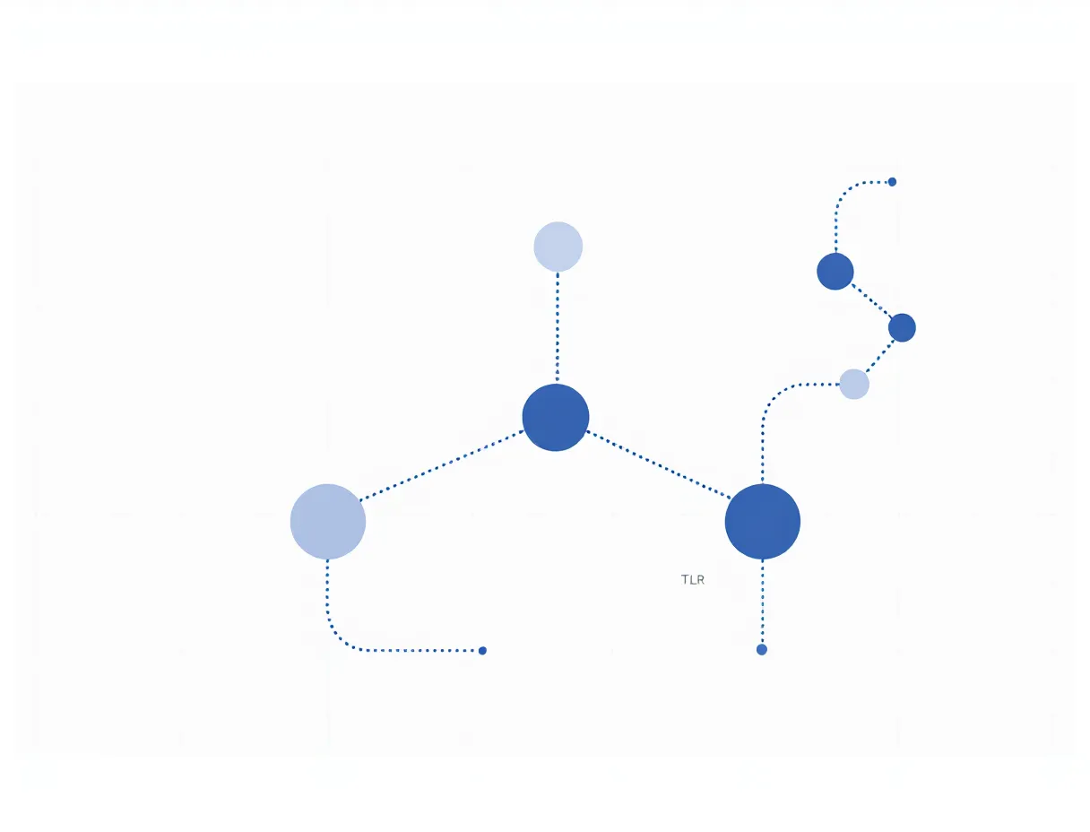
The primary mechanistic characterization of Tα1 comes from a 2004 study in Blood [5] that defined its action on dendritic cells via TLR2 and TLR9 signaling. Tα1 binds TLR2 and TLR9 on dendritic cells, triggering the p38 MAPK and NF-κB pathways. This drives functional maturation of the dendritic cells — upregulation of CD40, CD80, CD86, and MHC class II — and induces IL-12 production.
IL-12 is the pivotal cytokine here: it drives differentiation of naive CD4+ T cells into Th1 effectors secreting IFN-γ and IL-2. Downstream, CD8+ cytotoxic T lymphocytes expand, and natural killer cell activity increases. The net effect is a restoration of adaptive immune tone — specifically in settings where that tone is suppressed, whether by chronic infection, sepsis-induced immune paralysis, cancer, or age-related thymic involution.
In hyperinflammatory states, the response is different: Tα1 reduces proinflammatory cytokines including TNFα, IL-1β, and IL-6 rather than amplifying them. This bidirectionality — immunostimulatory in immunosuppressed states, immunomodulatory in hyperinflammatory states — is what distinguishes thymalfasin from a simple immune stimulant. The 2025 immunosenescence review confirmed this dual action: Tα1 increases IL-10 while reducing TNFα and IL-1β in aging models, and provides antioxidant protection to thymic epithelial cells [17].
In cancer, the additional IDO1 pathway has been characterized: Tα1 activates IDO1-dependent tolerogenic signaling, which in murine models protects against checkpoint inhibitor-induced colitis without impairing anti-tumor immune responses [9]. This mechanistic duality — T cell expansion in the tumor immune axis, gut mucosal protection against immune activation — is the biological rationale for the sequential melanoma studies.
Sepsis: ETASS, Meta-Analyses, and the TESTS Phase 3 Null Result
The sepsis literature is the most contested area in the Tα1 evidence record.
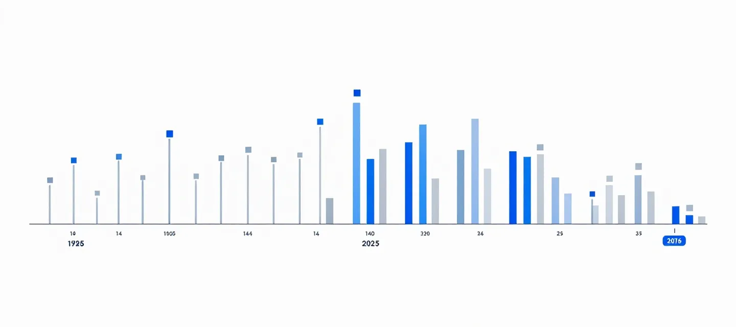
The ETASS trial (2013) [6] was a multicenter, single-blind RCT of 361 severe sepsis patients across six Chinese tertiary hospitals. Tα1 was administered at 1.6 mg subcutaneous twice daily for five days, then once daily for two days, alongside standard care. The 28-day mortality in the Tα1 arm was 26.0% versus 35.0% in the control arm — a 9 percentage point absolute reduction (log-rank P = 0.049). Monocyte HLA-DR expression, the pharmacodynamic marker for immune paralysis reversal, improved significantly at day 3 (+3.9%, P = 0.037) and day 7 (+5.8%, P = 0.017).
A 2016 systematic review and meta-analysis of 10 RCTs involving 530 sepsis patients [7] reported a pooled 28-day mortality risk ratio of 0.59 (95% CI 0.45–0.77; P = 0.0001). HLA-DR expression improved (SMD 1.23) and APACHE II disease severity scores were reduced with once-daily dosing. These results appeared confirmatory.
The TESTS trial (2025) [16] changed the picture. As the largest double-blind, randomized, placebo-controlled phase 3 trial of Tα1 for sepsis to date — 1,106 patients across 22 Chinese centers, enrolled 2016–2020 — it found no significant reduction in 28-day all-cause mortality: 23.4% in the Tα1 arm versus 24.1% in the placebo arm (HR 0.99, 95% CI 0.77–1.27; P = 0.93). Subgroup analyses identified trends toward potential benefit in elderly patients and those with diabetes; these findings are hypothesis-generating only.
A 2025 meta-analysis updating the sepsis evidence [15], incorporating 11 RCTs and 1,927 patients, found an overall 28-day mortality OR of 0.73 (95% CI 0.59–0.90; P = 0.003) — but when the analysis was restricted to high-quality multicenter trials, the benefit disappeared. The authors characterized the evidence base as heterogeneous and insufficient for definitive conclusions, recommending personalized immunotherapy trial designs targeting subpopulations with demonstrated benefit.
The current position of the Tα1 sepsis evidence: mechanistic plausibility is established and consistently replicated, the HLA-DR pharmacodynamic signal is robust, smaller and older trials showed mortality benefit, and the largest definitive phase 3 trial found no benefit. Both things are true. This site describes both.
Cancer: Checkpoint Inhibitor Synergy and Oncology Applications
The oncology data for Tα1 comes from two sources: retrospective clinical cohorts in metastatic melanoma, and a combination therapy review covering cancer, chemotherapy, and immunotherapy contexts.
In metastatic melanoma, retrospective data from patients who received sequential Tα1 followed by anti-CTLA-4 therapy showed a median overall survival of 38.4 months versus 8 months for anti-CTLA-4 monotherapy [9]. Long-term follow-up of 61 patients in this sequential cohort [10] extended the median OS estimate to 57.8 months — with a 5-year OS rate of 41.2% versus 13.0% in non-sequentially treated patients. These are striking numbers; they are also from a retrospective analysis with substantial selection bias potential. Prospective randomized confirmation does not yet exist.
The hypothesized mechanism involves two complementary effects: Tα1 expands the T cell pool prior to checkpoint release, potentially converting immunologically 'cold' tumors to immunologically active ones [9]; and Tα1 activates IDO1-dependent tolerogenic pathways that reduce checkpoint inhibitor-induced colitis, improving treatment tolerability and continuity.
In combination with chemotherapy, a 2000 review [11] documented that Tα1 combined with low-dose interferon or interleukin-2 restored tumor-growth-suppressed immune function and substantially reduced chemotherapy toxicity in both experimental and clinical settings.
In lung cancer, a 2025 retrospective study of 196 unresectable stage IIIA-IIIC NSCLC patients [14] found that weekly Tα1 for 12 months after concurrent chemoradiotherapy reduced grade ≥2 pneumonitis from 35.4% to 14.5%, reduced lymphopenia incidence from 55.8% to 22.5%, improved eligibility for consolidative immunotherapy (93.6% vs 75–77%), and suppressed IL-6. Median progression-free survival and overall survival were not reached in the long-term Tα1 arm at data cut-off. These are preliminary retrospective findings; prospective study is required before conclusions can be drawn.
A 2025 in vitro characterization study [18] examined Tα1's effects on tumor cell lines and immune cell subsets with important nuance. Direct transcriptional effects on tumor cells (melanoma, glioblastoma, mesothelioma) were minimal. The strongest effects were on activated CD8+ T cells; CD4+ T cells, B cells, and NK cells showed enhanced proliferation. Plasmacytoid dendritic cells upregulated CD40, CD80, TIM-3, and TNFα. However, CTL cytolytic activity against tumor targets was not enhanced, raising questions about direct anti-tumor immunomodulatory claims.

COVID-19, Severe Acute Pancreatitis, and Recent Findings
In COVID-19, a 2022 meta-analysis [13] of 9 studies involving 5,352 patients (1,152 receiving Tα1) found no statistically significant overall mortality reduction. Subgroup analyses identified potential benefit in patients over 60 years (RR 0.68) and in severe or critical COVID-19 cases (RR 0.66). The authors cautioned against broad use and called for confirmatory trials.
In severe acute pancreatitis (SAP), the evidence is more consistent. A 2025 meta-analysis [14] of 5 RCTs involving 706 SAP patients found Tα1 significantly increased CD4+ T cell percentages (MD +4.53), improved the CD4/CD8 ratio (MD +0.42), reduced extrapancreatic infection incidence (RR 0.56), reduced APACHE II scores (MD -1.52), and reduced C-reactive protein at lower doses. These results support an immune-regulatory benefit in the context of SAP's characteristic immune suppression following the initial hyperinflammatory phase.
In a double-blind pilot RCT of 24 severe acute pancreatitis patients using 3.2 mg Tα1 twice daily for 7 days [8], HLA-DR expression and CD4/CD8 ratios recovered more rapidly than in the control arm, and positive infection culture rates at day 28 were significantly lower. ICU duration was shorter in the treatment group.
The 2024 comprehensive review by Dinetz and Lee [12], which examined over 30 trials and 11,000 subjects across COVID-19, autoimmune disorders, and cancer, concluded that thymalfasin demonstrated consistent safety and efficacy as an immune modulator across indications, and characterized the FDA's 2023 Category 2 restriction as unfounded based on the clinical evidence. That characterization is a claim in the literature, not a regulatory conclusion.